|
PinkMonkey Online Study Guide-Biology
22.1 Pituitary Gland
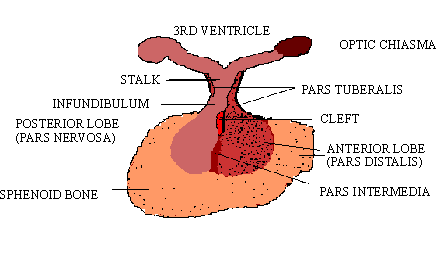
(A) Position: The pituitary gland (hypophysis) is about the size of a large pea, located below the brain, behind the nasal cavity on the floor of the cranium. It is attached by a stalk to the hypothalamus portion of the brain on the undersurface of the cerebrum (Figure 22.2). It lies in the sphenoid bone of the skull and is closely invested by a connective tissue capsule.
Click here for enlargement
Figure 22.2 Position of Pituitary gland
The pituitary has been called the "master
endocrine gland" because of its control over several other
endocrine glands. However, the bodys real master gland is
the hypothalamus of the brain which controls the secretions of the
anterior pituitary (by releasing and inhibiting hormones). Also,
the pituitary itself is in turn controlled by feedback from other
glands.
(B) Structure.
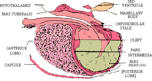
The sagittal section shows that the pituitary consists of two main lobes,
the nterior lobe (pars distalis) and the posterior
lobe (pars nervosa). The small intermediate lobe (pars
intermediate) is marked by a cleft (in children) or cystic
spaces (in adults) (Figure 22.3) The part of the anterior lobe which
extends upwards as part of pituitary stalk is called the pars tuberalis.
The pars anterior, pars tuberalis and pars intermedia form the
adenohypophysis while the pars nervosa and the pituitary stalk
( infundibulum) form the neurohypophysis. The anterior lobe produces
six different hormones. Of these, two work directly on body tissue (somatotrophic)
and the other four control the action of other endocrine organs. The
middle lobe (pars intermedia) secretes only one hormone, intermedin,
which controls skin colors in lower vertebrates, but is vestigial in mammals,
and has no function in humans. The posterior lobe serves as a storage
and release point for two hormones secreted by the hypothalamus (neurosecretory
cells) and carried to the lobe through a connecting duct (i.e., the neurohypophysis).
Click here for enlargement
Figure 22.3 Section of Pituitary
(C) Histology. Histological structure of the pituitary shows different types of cells in three different parts of the pituitary. The cells are classified as chromophobes or chromphils by the staining reaction of their cytoplasmic granules. (Figure 22.4) (Tripple Mellory stain method)
In the anterior lobe, chromophobe cells are smaller, nuclei clumped together and are clustered near the center. Chromophobe cells stain poorly. The chromophil cells stain red or purple blue, contain secretory granules and are distributed at the periphery. The nuclei differ in size and in staining intensity. They are of two types, oxyphilic cells and basophilic cells. Blood vessels and lymph spaces are also seen.
[next page]
|