|
PinkMonkey Online Study Guide-Biology
6.2 Mitosis
Mitosis is the characteristic division of the body cells,
hence called somatic division. It can be studied in the meristematic cells
in root and stem tips of plants.
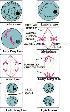
Figure 6.2 Various stages in the mitosis of a plant cell
Definition : "Mitosis is an equational division, dividing the mother cell into two daughter cells which are identical to each other and also to the original mother cell in every respect. In mitosis, the chromosomes of the mother cell are duplicated and distributed equally to the two daughter cells."
The mitotic cell cycle consists of interphase and M-phase.
Interphase is sub-divided into G1, S and G2 phases (as described
earlier in the cell cycle). The M-phase consists of karyokinesis
followed by cytokinesis.
Interphasic nucleus : The nucleus increases in volume during interphase. At this stage, the nuclear membrane and nucleolus are prominently visible. The chromosomes appear to form a continuous network (nuclear reticulum or chromatin network) of very fine threads. DNA replication has taken place (S-phase) and chromosomes have doubled. Interphase ends as the karyokinesis begins.
Karyokinesis : It involves a series of changes
within the nucleus. This is a continuous process but, for convenience,
it has been divided into four phases. These are (1) Prophase (2) Metaphase
(3) Anaphase and (4) Telophase. The main features of the nuclear division
during each phase are summarized below.
(1) Prophase : During early prophase, the chromatin
network becomes visible as it condenses into separate threads or chromosomes.
At this stage, each chromosome appears as a very fine, long single thread,
the chromonema and is it described as the monad. The nucleus envelope
and nuclear are prominently visible.
As the prophase progresses, chromosomes become shorter
and thicker (due to the condensing of their coils). In each chromosome,
the chromonema splits lengthwise into two identical threads or chromonemata
(dyads). These are coiled round one another. Chromosomes become more distinct.
Each chromonema becomes short, thick and is called a chromatid. At this
stage, each chromosome is shorter, thicker and consists of two identical
sister chromatids joined together by a spherical body called a centromere
(kinetochore).
By the end of prophase, the nuclear envelope and nucleolus
disappear completely. The chromosomes remain distributed in the nucleoplasm.
(2) Metaphase : Metaphase begins with the formation of a bipolar spindle body in the region of nucleoplasm (i.e. center of the cell). It consists of numerous spindle fibers. These are fine thread-like structures formed by the organization of proteins called tubulin into microtubules. There are two types of fibers in the spindle (a) Continuous fibers which extend from pole to pole and (b) Chromosomal fibers which extend from pole to the center (equator of the spindle).
The chromosomes move and get arranged in a plane along the equator of the spindle. This results in the formation of the equatorial plate (metaphasic plate). The centromere of each chromosome in the plate is connected with both the poles by the chromosomal fibers.
(3) Anaphase : During early anaphase, the centromere of each chromosome divides longitudinally into two. As a result, each chromosome is now completely divided into two identical halves (sister chromatids) called daughter chromosomes. The centromere of each daughter chromosome remains connected to the pole on its respective side by a chromosomal fiber.
During late anaphase, the two groups of daughter chromosomes are pulled away from each other and start moving towards the opposite poles. Most probably, this movement is caused by the shortening of the chromosomal fibers. In each group, chromosomes appear 'V' or 'L' shaped as the centromeres are pulled towards the poles and the chromosomal arms trail behind.
|