|
PinkMonkey Online Study Guide-Biology
(c) Anatomy of a sporophyll
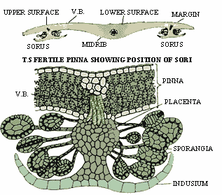
Figure 15.2 Transverse section of a fertile pinna through
the sorus
The transverse section of a fertile pinna passing through
a sorus shows the internal structure of the leaflet as well as that of
the sorus. The internal structure of the leaflet is differentiated into
upper and lower epidermis, spongy mesophyll and vascular bundles. Both
the upper and lower epidermis are single layered, non-green and covered
with cuticle. Stomata are generally present in the lower epidermis only.
Mesophyll tissue is spongy in nature. This is composed of thin walled,
green and loosely arranged cells. The vascular bundles of lateral veins
are seen in the mesophyll region. Each vascular bundle is concentric
and hadrocentric (i.e., it has xylem cells in the center surrounded
by the phloem cells).
The sorus is seen attached to the lower epidermis.
It consists of a placenta, numerous sporangia and indusium. The placenta
is a fertile tissue of colorless cells. It develops as a cylindrical out-growth
from the lower epidermis and bears sporangia and an indusium. Sporangia
are the spore producing organs. They develop laterally from the placenta.
The indusium is a thin membrane attached at the end of the placenta
which protects the sporangia in a young sorus.
|