|
PinkMonkey Online Study Guide-Biology
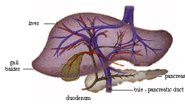 Figure 16.9 Liver and Pancreas
(b) Liver : It is the largest gland in the body, situated
in contact with the stomach. The human liver is imperfectly bilobed. Between
the lobes lies a green sac-like structure called the gall bladder
which stores the secretion of liver (called bile). The hepatic
duct from the liver lobes and the cystic duct from the gall bladder join
to form the common bile duct.
(c) Pancreas : It is an elongated lobulated gland situated
in the loop of the duodenum. The pancreatic duct joins the common
bile duct and opens into the duodenum.
Histology of the digestive system
The wall of the alimentary canal is made up of concentric
layers of serosa, (outermost), muscularis, sub-mucosa
and mucosa, (innermost). According to the function of the part,
they are modified as described below:
Histology of stomach: A transverse section of stomach
shows four concentric layers as described below :
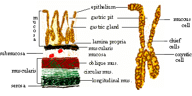
Figure 16.10 (A) Transverse
Section (B) Single gastric gland
(i) Serosa : It is the outermost single layer
of cells.
(ii) Muscularis : It is comparatively thick and distinguished into outer layer of longitudinal muscles, middle thick layer of circular muscles and inner layer of oblique muscles.
(iii) Sub-mucosa : It is a spongy layer situated between the mucosa and muscularis. It contains blood vessels and nerve endings. Between the sub-mucosa and mucosa, there is a distinct layer of muscularis mucosa.
[next page]
|