|
PinkMonkey Online Study Guide-Biology 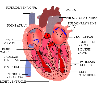
Click here to enlarge
Figure 18.6 Internal structure of the heart
The ventricles. The right and left ventricles
are separated by inter ventricular septum. The septum slopes
obliquely with convexity towards the right ventricle. The left ventricle
is longer and more conical than the right, and forms the apex
of the heart. The left ventricle shows the following
important features:
-
The left atrio-ventricular opening guarded
by a mitral or bicuspid valve.
-
A circular opening of the aorta guarded by
aortic or semilunar valves. The valves allow the blood
to enter the aorta from left ventricle during ventricular contraction
and prevents it from flowing back into the ventricle during
relaxation. The aortic valve has 3 cups-two posterior (right
and left) and one anterior.
-
The trabeculae carneae (chordae tendinae),
are attached to the margins of the bicuspid valve and prevent
them from everting into the atrium.
-
The papillary muscles, two in number, to which
one end of the chordae tendinae is attached .
The right ventricle has :
-
Right atrio-ventricular opening guarded by
the tricuspid valve.
-
A rounded opening of the pulmonary artery,
guarded by semilunar valves. The semilunar valves prevent backflow
of the blood.
-
The trabeculae carneae are as in the left ventricle,
but they are not as stout and strong.
-
The papillary muscles are conical in shape
with their bases attached to the walls of the ventricle and
their apices directed towards the ventricular cavity.
[next page]
|
Table of Contents
18.0 -
Introduction
18.1 -
Closed Vascular System
18.2 -
Heart
18.3 -
Arterial Blood Pressure
18.4 -
Blood
Chapter
19
|