As the blastocyst reaches the uterus, it attaches
and sinks in the uterine endometrium. This is called implantation
and occurs 7-8 days following fertilization. During implantation,
the trophoblast cells secrete enzymes which help the blastocyst
to attach and implant. The normal site of implantation is in the
posterior wall of the uterus. Implantation helps the blastocyst
to absorb nutrients from the glands and blood vessels of the endometrium
for its growth and development.
Gastrulation
Gastrulation is defined as a dynamic process during
which rearrangement and reorganization of the cells take place to
form the three primary germ layers: the ectoderm, endoderm and the
mesoderm.
In humans, the formation of the germ layers happens
so quickly that it is difficult to determine the exact sequence
of events.
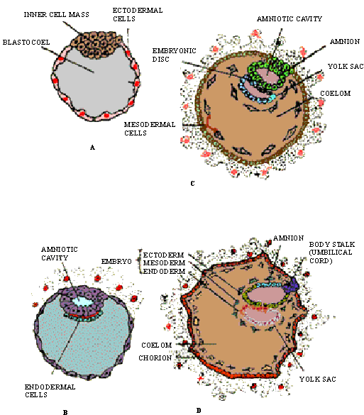
(1) Before implantation, a layer of ectoderm
(the trophoblast layer) has been formed around the blastocoel
(Figure 24.12A). The trophoblast cells will become part of the chorion
#one of the membranes surrounding the fetus.
(2) The inner cell mass moves downwards,
and a space called the amniotic cavity is formed within the
inner cell mass. The bottom layer of the inner cell mass develops
into an endodermal germ layer (Figure 24.12B)
(3) Now a layer of cells grows around the
top of the amniotic cavity, to form the amnion-another fetal
membrane. The cells below the cavity form the embryonic disc,
from which the embryo develops. The embryonic disc contains scattered
ecto, meso and endodermal cells.
(4) The endodermal cells now divide rapidly
and extend downward in a circle (Figure 24.12C). In birds and reptiles
this circle is the yolk-sac yet another fetal membrane. In
mammals, an allantoic membrane also forms from endosperm, which
stores metabolic wastes in some species. Some of the mesodermal
cells also move into the structures of the fetal membrane.
(5) The embryonic disc separates into 3
distinct layers: The upper ectoderm, the middle mesoderm,
and the lower endoderm (Figure 24.12D). The embryonic disc gradually
squeezes off the yolk-sac, and the cavity inside the disc
is the endoderm-lined primitive gut. The mesoderm within
the disc soon splits into an outer somatic and inner splanchnic
mesoderm. The space between these layers becomes the coelom,
or body cavity.