|
|
|
(B) Histology of kidney: The vertical section
of a kidney (Fig.19.2) shows that it consists of two regions, the
outer dark region called the cortex, and the inner, lighter colored
zona medulla. It also shows a large number of tiny tubules (nephrons),
many capillaries and connective tissue. There is a collecting space
called the pelvis where the ureter leaves the kidney; 6 to 15 cones
or pyramids of kidney tissue project into this space.
The renal artery divides into capillaries, which
carry blood to the glomerulus of the uriniferous tubules. The
renal vein carries blood away from the uriniferous tubules through
its capillary network.
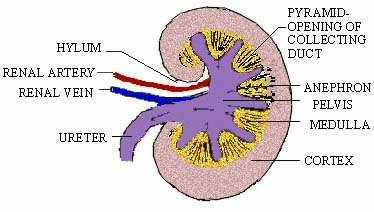
Figure 19.2 Sagittal Section of a Kidney
i) Structure of a uriniferous tubule (Nephron)
The nephron (Fig. 19.3) is the basic excretory
unit of the kidney; there are over a million in each kidney. Each
nephron consists of a glomerulus, Bowmans capsule and
associated renal tubules.
The glomerulus is a small knot of blood vessels
formed by a capillary network from the renal artery (afferent vessel).
The smaller efferent vessels take the blood away from the glomerulus
and enter the capillary network around the tubule of the nephron.
The capillaries unite to form the venules to form the renal vein
which joins the inferior vena cava.
|
[next page]
|
Table of Contents
19.0
- Introduction
19.1 - Ammonotelism, Ureotelism and Uricotelism
19.2 - Excretory System of Man
19.3 - Skin and Lungs as Accessory Excretory Organs
Chapter 20
|